Cat Ultrasound, MRI, XRAY and Radiology
 VETERINARY DIAGNOSTIC IMAGING FOR CATS
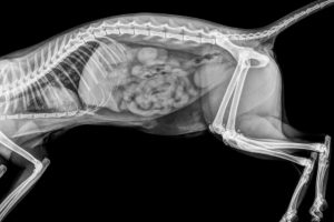
Veterinary diagnostic imaging includes modalities such as radiographs (x-rays), ultrasound, MRIs, and CT scans, which are used to gather information on the appearance and function of internal structures in a cat’s body.
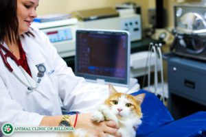
While these modalities are non-invasive and painless, some imaging requires sedation or general anesthesia because the cat must remain very still for a period of time to allow for the collection of clear images.
Often, diagnostic imaging provides essential information about a disease process or injury. The images collected are used by your veterinarian, along with physical exam findings and other diagnostic tests, to diagnose your cat’s condition and create a treatment plan.
WHY VETERINARY DIAGNOSTIC IMAGING IS NECESSARY?
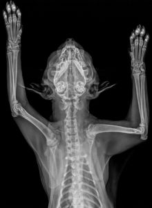
Radiography (x-ray) is by far the most commonly-used form of veterinary diagnostic imaging, and is usually employed before other forms of imaging. X-rays may either reveal a diagnosis (such a broken bone or bladder stones), or may furnish important information to direct further testing (such as an enlarged liver that may have several possible causes).
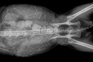
An ultrasound exam will often furnish information that is complimentary to the information gathered from x-rays. In some situations, advanced imaging—either a CT Scan or an MRI—may be necessary to adequately visualize an abnormality. An example of this would be a lesion in the brain or spinal cord, for which MRI produces the most detailed images.
Veterinary diagnostic imaging helps our veterinarians make a sound diagnosis of your cat’s condition. Imaging equipment includes:
- X-Rays
- Ultrasounds
- MRI’s
- CT’s
More information on each of these types of radiographs is provided below.
CAT X-RAYS
X-rays have been used by the medical community for over a century. They are the most regularly used form of diagnostic imaging in the veterinary industry because they are cost-effective and provide a great deal of information. X-rays are painless, but some cats benefit from sedation to reduce anxiety. Modern x-ray equipment generates relatively low levels of radiation that is very safe for the patient.
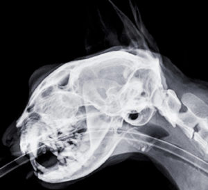
Taking cat x-rays:
- The x-ray machine settings are adjusted to produce the best image based on the thickness and density of the area being imaged
- The cat is precisely positioned on the x-ray table to allow for the best possible view of the area of interest
- The x-ray machine is positioned so the beam will target the area of interest
- Each x-ray takes only a split second to generate, and with digital x-ray technology, each image is available for review almost instantly.
- Digital x-ray technology also allows for rapid and effortless storage and transfer of x-ray images.
X-rays can provide a great deal of information, including:
- Orthopedic problems: abnormalities of the bones, joints, and surrounding structures.
- Heart problems: changes to the size and shape of the heart and associated blood vessels are often characteristic of specific disease processes
- Lung disease: infiltration of tissue or fluid into specific areas of the lungs can reveal the presence of pneumonia, asthma, cancer, and other diseases.
- GI Tract abnormalities: many ailments of the esophagus, stomach, and intestines can be detected with x-rays, including intestinal blockages, entrapped foreign material, and constipation/megacolon
- Liver, spleen, and kidney issues: changes to the sizes and contours of these organs on x-rays can indicate the presence of disease
- Kidney and bladder stones: Most types of kidney and bladder stones are visible on x-rays
- Detection of enlarged lymph nodes and masses
- Dental and periodontal disease: dental x-rays are an integral part of a complete dental evaluation and cleaning; because the majority of each tooth is below the gum line, problems such as broken roots and tooth root abscesses can only be detected using x-rays
CAT ULTRASOUNDS
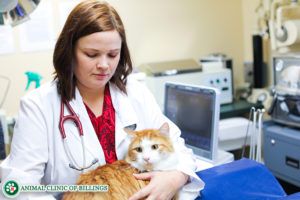
An ultrasound is the second most common diagnostic imaging tool veterinarians use to learn more about a cat’s condition.
Ultrasound uses sound waves to visualize the internal composition of different organs and tissues, and can also be used to evaluate dynamic processes in real time, such as the opening and closing of the heart valves and the movement of the intestines.
Performing a cat ultrasound:
 The cat is gently positioned on his or her back in a soft foam trough; for some patients, a mild sedative may be given to reduce stress and movement. The fur over the area to be imaged is clipped to allow for good contact between the ultrasound probe and the skin surface. Either alcohol or ultrasound gel is applied to the area to optimize sound wave transmission and produce the clearest possible images. The ultrasound probe, which emits a beam of ultrasound waves, is placed in contact with the skin; as these waves encounter tissues of varying densities they are reflected back to the probe, generating an image of the underlying body part on the ultrasound screen.
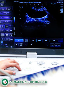 By sliding the probe around and changing the direction and angle of the beam, an area can be thoroughly visualized. Still images and short video clips can be captured for storage and further review, including precise measurements. Doppler technology can be used during an ultrasound exam to visualize blood flow through the heart and larger blood vessels.
Ultrasound can be used to evaluate:
- Abdominal organs and structures
- The heart and surrounding structures
- Lymph nodes and glands
- Muscles, tendons, and ligaments
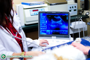 Ultrasound also enables a veterinarian to precisely guide a needle into a tissue or fluid to collect a sample for microscopic analysis.
Because ultrasound and x-rays often provide complementary information, these two modalities are often used together in diagnosing an illness.
For example, in the evaluation of heart disease, x-rays reveal any changes to the size and shape of the heart and great vessels, while ultrasound provides information about cardiac wall thickness, chamber size, and valve function.
 MRI’s FOR CATS
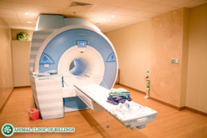
Magnetic resonance imaging (MRI) is the newest form of diagnostic imaging in both human and veterinary medicine.
In an MRI scan, a combination of a powerful magnetic field and radio waves is used to produce detailed images of a cat’s internal anatomy.
MRI scans are completely painless and very safe, involving no radiation exposure.
A cat MRI involves the following steps:
- An IV catheter is placed in the cat’s front leg for the administration of sedatives and contrast solution
- Bloodwork is checked to ensure there are no problems that would make sedation, anesthesia, or contrast administration dangerous
- The cat is heavily sedated or anesthetized so they will remain still for the duration of the scan
- The cat is positioned on a padded gurney that slides into the opening in the center of the MRI scanner
- The scan may take anywhere from 15 to 60 minutes, depending on how much of the body is being scanned
- Often, an IV injection of a contrast solution will be administered partway through the scan to provide detailed images of blood flow within an area of interest
- Following the MRI, the cat will remain on IV fluids for at least a few hours as the sedation or anesthesia wears off
- The MRI images are transmitted to a radiologist who examines them closely and sends back a report detailing their findings
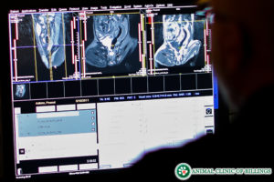 Due to their cost and the specialized equipment and expertise required, cat MRI’s are usually only performed in cases where x-rays and ultrasound are not sufficient to determine a diagnosis.
CT SCANS FOR CATS
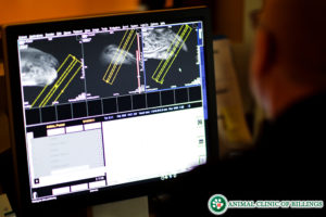
CT scans for cats, also called “cat scans,” use x-rays to generate detailed three-dimensional images of internal anatomy. While they do not provide as much detailed information about the status of soft tissues as an MRI does, they are excellent for evaluating bone, and do provide valuable information about the body’s soft tissue structures.
They are most commonly used to evaluate bone and joint abnormalities, the lungs and associated structures, and the abdominal organs. CT scans have the advantage of being much more rapid to perform; typically only requiring a few minutes. Even though the images are acquired more rapidly, it remains essential that the patient remain very still throughout the scan, so brief sedation or anesthesia is required.
A cat CT scan involves the following steps:
- An IV catheter is placed in the cat’s front leg for the administration of sedatives and contrast solution
- Bloodwork is checked to ensure there are no problems that would make sedation, anesthesia, or contrast administration dangerous
- The cat is heavily sedated or anesthetized so they will remain still for the duration of the scan
- The cat is positioned on a padded gurney that advances slowly through the center of the CT scanner while the rotating x-ray beam passes through the body part, allowing the rotating detector to collect detailed information about tissue density from all directions.
- An IV injection of a contrast solution may be administered to highlight blood vessels within an area of interest
- Computer processing transforms the collected data into detailed cross-sectional images of the area under study
- The scan typically takes only 5-10 minutes
- Following the CT scan, the cat will remain closely monitored until the sedation or anesthesia wears off
- The CT images are transmitted to a radiologist who examines them closely and sends back a report detailing their findings
As with MRI’s, CT scans are more costly and require specialized equipment and expertise. CT scans are usually only performed in cases where x-rays and ultrasound are not sufficient to determine a diagnosis.
 The Animal Clinic of Billings and Animal Surgery Clinic is here to help with all your cat care needs.
If you are concerned that your cat might be injured or experiencing internal problems, or to discuss how feline radiographs can benefit him or her, please contact us to schedule an appointment with one of our veterinarians today.
406-252-9499
ANIMAL CLINIC OF BILLINGS AND ANIMAL SURGERY CLINIC
providing our region’s companion animals and their families what they need and deserve since 1981
1414 10th St. West, Billings MT 59102
406-252-9499


